
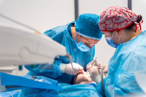
Some lower teeth sit close to nerves that give feeling to your lower lip, chin, teeth, gums, and tongue. Lower molars and wisdom teeth are the most common examples. Nerve protection during tooth extraction starts with knowing where those nerves run before surgery begins.
Two nerves matter most. The inferior alveolar nerve (IAN) runs through a canal inside the lower jaw. The lingual nerve travels through soft tissue on the tongue side of the jaw. Closeness to either nerve does not mean an injury will happen. It tells the oral surgeon to evaluate the anatomy carefully and adapt the approach when needed. The Oral Surgery Group plans each surgical tooth extraction around this anatomy for patients across Bucks County, Montgomery County, and the greater Philadelphia region.
Two sensory nerves run near the lower back teeth, and a third branch carries feeling to the lip and chin. Knowing what each one does explains why lower extractions get such careful planning.
The inferior alveolar nerve is a branch of the mandibular division of the trigeminal nerve. It enters the lower jaw near the back and travels forward through a bony tunnel called the mandibular canal.
Along the way, it supplies sensation to the lower teeth, gums, and tooth sockets. A StatPearls anatomy review notes that it also supplies the front of the chin and the lower lip through the mental nerve.
The roots of some lower molars sit close to this canal. In some patients, only a thin layer of bone separates the two.
The lingual nerve runs through the soft tissue along the inner, tongue-facing side of the lower jaw. Near the wisdom teeth, it can lie close to the bone and gum tissue.
This nerve carries general sensation from the front two-thirds of the tongue, the floor of the mouth, and the inner gums. It also carries taste fibers from the front of the tongue through its connection with the chorda tympani.
Unlike the IAN, the lingual nerve does not run inside a bony canal. That makes it a soft-tissue concern rather than something surgeons trace through the jawbone.
The mental nerve is a terminal branch of the inferior alveolar nerve. It exits the jaw through a small opening called the mental foramen, usually below the premolars.
It supplies the lower lip and chin. Because it continues from the IAN, an injury farther back along the nerve can show up as lip or chin numbness.
The back of the lower jaw packs several risk factors into a small area.
Not every lower molar sits near both nerves. Many lie well away from either one, and imaging shows where a specific tooth falls.
Sitting near a nerve and injuring a nerve are two different things. A nerve can lie close to a root and come through surgery unaffected. When changes do occur, they trace back to a few general mechanisms.
It can. Some roots lie extremely close to the mandibular canal. Others contact or groove the canal wall, which surgeons describe as an intimate relationship.
Imaging helps assess this relationship. A panoramic X-ray cannot always confirm true contact, though. A root can overlap the canal on a flat image while sitting beside it in three dimensions.
Nerve changes after extraction generally stem from one of several mechanisms.
Each mechanism affects the nerve differently. That difference shapes how sensation may recover.
Yes. Nerve injury exists on a spectrum. A mild injury may only disrupt how signals travel for a time. A more serious injury damages the nerve fibers themselves, and complete cutting sits at the far end.
Some sensory changes fade as the nerve recovers. Others can persist.
Every root leaves the socket along a path. If that path drives a root toward the canal, it can press on the nerve.
Root orientation, curvature, and surgical access all influence which direction keeps pressure away. For that reason, the surgeon plans the removal path for each tooth individually.
No single feature predicts nerve injury. Surgeons weigh several factors together to build a picture of risk for each tooth.
The closer the roots sit to the canal, the more detailed the evaluation becomes. Close proximity calls for careful imaging review and a deliberate removal plan. It does not, by itself, mean an injury will happen.
Impacted lower wisdom teeth bring several variables at once.
A deep, angled tooth with mature roots near the canal needs more planning than a partly erupted tooth with short roots.
Yes. Curved or spreading roots may not follow a straight path out of the socket. Near the nerve, that detail matters.
A root that hooks toward the canal may need a different removal path than a straight one.
They can. Bone can become denser and less flexible with age, and roots finish developing in early adulthood. Both can make removal more involved.
Neither factor predicts nerve injury on its own. Risk depends on how several factors combine for a given tooth. Other jawbone changes can also shift the plan, as explained in how bone loss changes tooth extraction planning.
Past problems can change the anatomy a surgeon works with.
The surgeon reviews this history alongside the imaging.
Planning combines imaging, a clinical exam, and a frank conversation with you. Each part answers a different question about the tooth.
A panoramic X-ray shows the entire lower jaw in one image. It helps surgeons judge the approximate relationship between molar roots and the mandibular canal.
Certain findings suggest a closer relationship.
These signs call for closer review. A panoramic image cannot always confirm whether a root directly touches the nerve.
Cone-beam computed tomography (CBCT) creates a three-dimensional view of the jaw. Surgeons may recommend it when a panoramic X-ray shows a close or uncertain relationship and the answer could change the plan. A CBCT scan can show several details.
CBCT is not necessary for every extraction, and it does not eliminate nerve risk. A 3D scan shows the anatomy more clearly, but it cannot change where the nerve sits. Its value lies in clarifying that anatomy for planning.
Routine dental X-rays do not show the lingual nerve. Its exact path also varies between people.
Surgeons account for it using known anatomical pathways, the clinical exam, and what they see during surgery. The surgeon plans access to the tooth with that pathway in mind.
An exam captures details an image cannot.
Recording existing sensory changes before surgery also creates a baseline for comparison afterward.
Informed consent covers why the tooth needs treatment and what removal should accomplish. It also covers the realistic sensory risks for your anatomy and any alternatives, such as monitoring or partial removal.
No surgeon can promise zero risk. A clear conversation helps you weigh the options with accurate expectations.
The goal is to remove the tooth with as little force near the nerve as possible. A few principles guide that work.
Root position, surrounding bone, and nearby structures all shape the approach. A canal on the tongue side of the roots calls for a different plan than one sitting directly beneath them.
The surgeon chooses access and removal paths that keep pressure away from the canal.
Controlled removal follows a few habits.
The same principles explain why some teeth require multi-piece removal.
Sectioning divides a tooth so each portion can come out separately. Near the nerve, this can let the surgeon remove smaller pieces along safer paths.
Sectioning does not lower nerve risk in every case. Whether it helps depends on the tooth. A separate guide explains tooth sectioning during extraction in more detail.
On the inner side of the jaw, the lingual nerve sits in soft tissue close to where surgeons work on lower wisdom teeth. Gentle handling of that tissue limits pressure and stretching on the nerve.
Yes. Several options remain open.
Sometimes the surgeon recommends monitoring a symptom-free, high-risk tooth instead of removing it.
A coronectomy removes the crown of a tooth while intentionally leaving selected roots in place. Surgeons may consider it for certain impacted lower wisdom teeth whose roots sit very close to the canal.
In carefully selected cases, coronectomy offers an alternative to complete extraction that can lower the risk of injury to the inferior alveolar nerve.
Complete extraction removes the crown and every root. Coronectomy removes the crown and leaves selected roots beneath the bone.
It is a specialized option for specific high-risk teeth. It is not routine treatment for molars.
Roots closely tied to the canal carry the most risk during removal. Leaving them in place avoids manipulating them next to the nerve. In appropriately selected cases, this may lower the chance of an IAN injury.
Suitability depends on clinical and radiographic findings. The condition of the tooth and roots matters, as does any related disease.
Surgeons also consider whether the retained roots can stay healthy under the bone. An individual evaluation determines whether coronectomy fits a particular tooth.
Retained roots can shift position over time, a change called root migration. In some cases, a root moves closer to the surface of the gum or bone. A small proportion of patients need a second procedure, often because a root becomes exposed. Regular follow-up visits and imaging help the surgeon monitor the retained roots over time.
Some numbness after extraction is normal. The real question is whether a change outlasts the anesthetic.
Yes. Local anesthetic numbs the lip, tongue, cheek, and nearby tissue on purpose. How long that lasts depends on the anesthetic used. Your surgeon will tell you roughly when feeling should return.
These changes usually appear on the same side as the treated tooth.
Lingual nerve changes affect the tongue on the treated side.
Yes. Paresthesia describes altered sensation, such as tingling, that is not painful. Dysesthesia describes altered sensation that feels unpleasant or painful, such as burning.
Some people notice increased sensitivity instead of reduced feeling. Others lose feeling in the area completely.
Call promptly if numbness or altered sensation lasts beyond the expected anesthetic period. The same applies if symptoms worsen or simply concern you.
Do not wait weeks to see whether it improves. Early reporting lets the surgeon document the change and plan follow-up.
Not always. Many sensory changes improve over time, and some do not. The outcome depends on the type and severity of the injury.
Many nerve-related changes after extraction are temporary. Recovery for any individual still varies. Recovery for any individual still varies.
Often, yes. A nerve irritated by bruising, stretching, or swelling may regain normal function as the tissue heals. Recovery depends on how much damage the nerve fibers sustained.
More severe injuries damage the nerve fibers themselves. Those fibers regrow slowly, and the pace varies between people and between nerves.
No one can give a precise recovery timeline at the outset. Follow-up visits track progress instead.
Yes. Persistent numbness or altered sensation is possible. The risk varies with the tooth, the procedure, and the anatomy involved.
This possibility is the reason surgeons discuss nerve proximity before surgery rather than after.
Evaluation builds a picture of the change over time.
No single treatment guarantees a return to normal sensation. Early evaluation gives the care team the most information to guide next steps.
No. Risk depends on which tooth is coming out, where its roots sit, and how far they are from the canal. Surgical complexity, individual anatomy, and other clinical factors also play a part.
They are the most common setting for nerve risk assessment. Impacted lower wisdom teeth often sit deeper and closer to both nerves.
The relationship still varies a great deal. Some wisdom teeth sit nowhere near either nerve.
Yes. A first or second molar can have long roots that reach toward the canal. When imaging shows that relationship, the surgeon plans for it like any higher-risk tooth.
No. Proximity shapes the risk assessment and the surgical plan. It does not automatically rule out extraction.
Instead, it guides the choice between complete removal, an alternative approach, or monitoring.
General dentists remove many teeth safely every day. Some cases benefit from a specialist’s focus on surgical anatomy, particularly when a nerve sits nearby.
Referral makes sense in several situations.
Oral and maxillofacial surgeons complete hospital-based surgical residency training after dental school. That training centers on surgical anatomy, diagnosis, and conditions affecting the mouth, jaws, and face.
It prepares them to plan around nerves, interpret 3D imaging, and offer alternatives when standard removal carries too much risk.
A few direct questions can make the conversation more useful.
Occasionally, yes. Removal can affect a nearby sensory nerve, most often with lower molars and wisdom teeth. Careful planning helps reduce that risk, and many sensory changes are temporary.
Both the inferior alveolar and lingual nerves. The inferior alveolar nerve affects lip and chin sensation, while the lingual nerve affects the tongue. Surgeons assess the first mainly with imaging and the second through anatomy and examination.
Yes, for the inferior alveolar nerve. Dental imaging helps assess how close roots sit to the mandibular canal. A flat X-ray cannot always confirm direct contact, so some cases need a 3D scan.
No. CBCT provides three-dimensional information that supports surgical planning. It does not guarantee prevention, because the nerve’s position and the surgery itself still carry risk.
It can feel like numbness, tingling, pins-and-needles, or a burning or unusual sensation. The change typically affects one side of the lip, chin, or tongue.
No. Numbness from local anesthetic wears off as the medication clears. Nerve-related numbness may be temporary or persistent depending on its cause and severity.
A coronectomy removes a tooth’s crown and intentionally leaves selected roots in place. It may suit certain impacted lower wisdom teeth whose roots sit very close to the inferior alveolar nerve.
Often, yes, after an individualized assessment. The surgeon weighs the risks of full removal against alternatives such as coronectomy or monitoring.
Yes. Persistent altered sensation warrants a prompt call. Note which areas feel different and when you first noticed the change, since those details help the surgeon assess it.
A tooth close to a nerve deserves a careful look before it comes out. The right plan starts with understanding exactly where the roots and nerves sit.
The Oral Surgery Group evaluates complex extractions for patients across Bucks County, Montgomery County, and the greater Philadelphia region. At the Doylestown or Meadowbrook office, your surgeon reviews how the roots relate to nearby nerves and builds an individualized surgical plan.
You’ll also get a clear discussion of risks and alternatives, plus guidance on what to expect and what to report after surgery.
Schedule a tooth extraction evaluation to talk through your options with an oral surgeon.


I had 2 molars removed (17 & 18) and didn’t feel a thing! Before I knew it, they were out! If I ever need oral surgery again there is no one else I will see. I would HIGHLY recommend him!
My doctor was great and the staff were great also. I would definitely recommend them to anyone. They were all very friendly and helpful.
I was able to get scheduled very soon after I first called in. And once I arrived at the office, the entire process from start to finish was prompt, professional, and expertly done. I am happy to recommend this practice to anyone.
Absolutely! I felt very confident with him. I will always return to him for any future dental work I need done. The whole experience was very well organized. I didn’t think a doctor and staff as good as this existed.
Dr. Dachowski is an amazing oral surgeon who is an expert in his field. I have had several implants placed by him and was a painless procedure. His entire team is professional and extremely concerned about their patients comfort and excellent care.
I traveled from Wilmington, DE because Dr. Dachowski and his staff are the best for teeth extractions and implants!
I am very happy with Dr. Dachowski and all of his staff. They have taken a displeasurable situation and made it the best it could be. My wife is also a patient and recommended me. I am very glad to have found them and would highly recommend them. As a business owner I understand the value of great customer service. Keep up the good work.
They are a very professional and impressive organization that uses the latest technology to do the job perfectly. They also take time to answer any questions you might have about the procedure. Will use them again and recommend them to others.
I highly recommend this practice for their quality service, knowledge & professionalism!